Zacharias Jansen and the first compound microscope
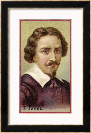
Then, during the 1590's, two Dutch spectacle makers, Zacharias Jansen and his father Hans started experimenting with these lenses. They put several lenses in a tube and made a very important discovery. The object near the end of the tube appeared to be greatly enlarged, much larger than any simple magnifying glass could achieve by itself.
Their first microscopes were more of a novelty than a scientific tool since maximum magnification was only around 9x and the images were somewhat blurry. Although no Jansen microscopes survived, an instrument made for Dutch royalty was described as being composed of "3 sliding tubes, measuring 18 inches long when fully extended, and two inches in diameter". The microscope was said to have a magnification of 3x when fully closed, and 9x when fully extended.
Although ordinary magnifying glasses are basically a simple microscope, when we speak of the invention of the microscope, we really mean the "compound microscope". Compound microscopes feature two or more lenses, connected by a hollow cylinder (tube). The top lens, the one people look through, is called the eyepiece. The bottom lens is known as the objective lens. So today, when we say "microscope," we really mean "compound microscope".
There is a lens called "the objective" which produces a primary magnified image. Then there is another lens called "the eyepiece" or "ocular," which magnifies that first image. In actual practice, there are several lenses used for both the objective and ocular, but the principle is that of two-stage magnification.
It is believed that Zacharias Jansen's father, Hans, helped him build the first microscope in 1595. Zacharias wrote to a Dutch diplomat, William Boreel, about the invention. When the physician of the French king inquired about the invention in the 1650's, Boreel recounted the design of the microscope.
Anton van Leeuwenhoek
It was Anton van Leeuwenhoek (1632-1723), a Dutch draper and scientist, and one of the pioneers of microscopy who in the late 17th century became the first man to make and use a real microscope.
Van Leeuwenhoek achieved greater success than his contemporaries by developing ways to make superior lenses, grinding and polishing five hundred and fifty lenses to make his new lens tube that had a magnifying power of 270x and could view objects one millionth of a meter (other microscopes of the time were lucky to achieve 50x magnification).
Van Leeuwenhoek made many biological discoveries using his microscopes. He was the first to see and describe bacteria, yeast plants, the teeming life in a drop of water, and the circulation of blood corpuscles in capillaries. During a long life he used his lenses to make pioneer studies on an extraordinary variety of things, both living and non living, and reported his findings in over a hundred letters to the Royal Society of England and the French Academy.
Van Leewenhoek's work was verified and further developed by English scientist Robert Hooke, who published the first work of microscopic studies, Micrographia, in 1665. Robert Hooke's detailed studies furthered study in the field of microbiology in England and advanced biological science as a whole.
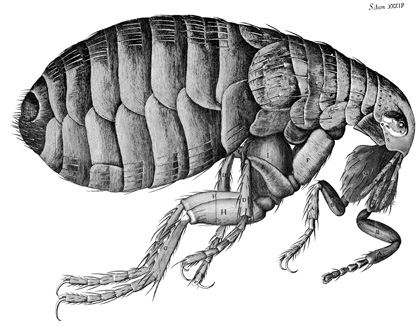
Hooke's Micrographia
Robert Hooke published Micrographia in 1665. It is his most famous work and is notable for the stunning illustrations, drawn by Hooke himself. Microphagia presents several accounts of Hooke's observations through the use of the microscope. He looked at all sorts of things (snow, a needle, a razor, etc.) with a primitive compound microscope, but his most significant observations were done on fleas and cork. He observed the fleas under the microscope and was able to observe the tiny hairs on the fleas' bodies. On the cork he saw pores. Upon examination of the pores, he decided to call them "cells"; however, he did not know he had just discovered plant cells.
Despite these great achievements in microscopy, microscopes didn't change much over the next 200 years, even though there were imperfections when viewing an object due to the different refraction of light. In the 1850s, German engineer Carl Zeiss began making refinements to the lenses he used in the microscopes he manufactured. In the 1880s, Zeiss hired glass specialist Otto Schott, who conducted research on optical glass, greatly contributing to the improvement of the optical quality of the microscope.
We should also mention Ernst Abbe, who was hired by Zeiss to improve the manufacturing process of optical instruments, which back then was largely based on trial and error. In a long and fruitful collaboration, Abbe carried out theoretical studies of optical principles, improving the understanding of the optical quality of a microscope.
Modern compound microscopes
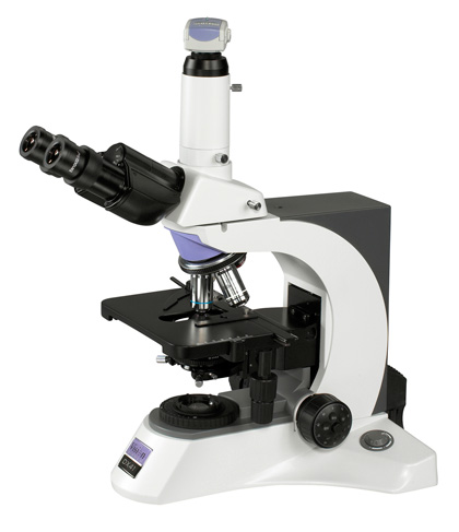
With the advancement of technology and improved optics, the microscope as we know it today came into being.
The theoretical minimum size able to be viewed by an optical microscope is 200nm (as defined by Abbe), since optical microscopes are only able focus on objects that are at least the size of a wavelength of light (usually, a wavelength of around 550 nm is assumed).
An electron microscope, in contrast, can magnify images thousands of times smaller than a wavelength of light.
